Ascites ultrasound is one of the tools for diagnosing ascites. Usually, the doctor would start by taking your medical history. He will ask a few questions, and sometimes a lot too, to detect risk factors. The medical history would also include questions about how you feel and if there are any symptoms. After this, the doctor will conduct a physical exam by checking your body for signs of ascites The shape of your belly while standing up and lying down can tell if you have ascites or not. Then gastrointestinal radiology tests such as ultrasound and/or CT scans may follow. If things are still not clear after radiology tests, your doctor may order paracentesis. The process may be quite long. But then, it is very vital to be sure about the diagnosis, as well as to detect the cause of ascites.
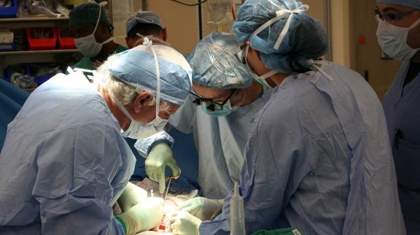
Paracentesis is like the final procedure to examine ascites. It is the next step after gastrointestinal (GI) radiology. It is an invasive procedure, so the doctor would wait for results of ultrasound and/or CT scan before deciding whether paracentesis may not be necessary. If the diagnosis is clear after GI radiology, you may not need paracentesis. But if the diagnosis is still not clear after GI radiology, which sometimes occurs, then you may need paracentesis. During paracentesis, lab tests will examine the ascitic fluid to know if there are signs of any underlying medical problem such as infection and cancer.
Gastrointestinal Radiology
Imaging tests are very vital diagnostic tools. They could help diagnose many diseases, including ascites. The radiographic features of ascites vary by the type of imaging test you are doing. More so, some of these tests are more sensitive than others.
A plain radiograph can help detect ascites. But then, the fluid in your peritoneum must be 500 mL at least before plain radiograph can detect it. If there is that us fluid in your peritoneum, a plain radiograph will show a general increase in the density of your abdomen.
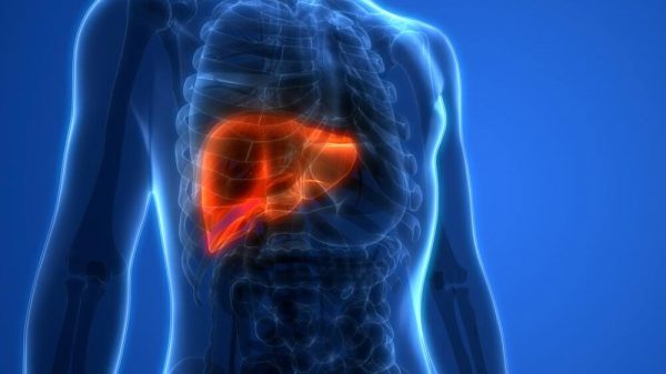
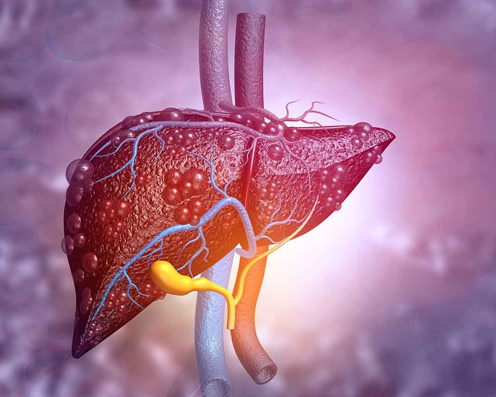
Beyond the density increase, the shadows of some of your soft tissues will be poorly defined if you have ascites. Examples of such soft tissues include your liver, spleen, and psoas muscles.
Furthermore, your solid visceral organs and your bowel will displace towards the midline of your belly. You flank would also bulge out.
Ultrasound, on the other hand, can detect smaller fluid volumes. If the fluid is adjacent to your diaphragm, then ultrasound is the best tool to detect it. Such is what happens in ascites.
Aside from detecting the presence of ascitic fluid, doctors can also assess the fluid with an ultrasound test. Simple ascites usually has no echo.
Hemorrhagic, exudative, or malignant ascites, on the other hand, usually contains some debris floating around. More so, if an ultrasound test shows septations, then there is likely to be inflammation or cancer.
CT scans can also detect very small fluid quantities in your peritoneum. It is more sensitive for fluid that lodges in the dependent regions like your pelvis and Morison pouch.
A CT scan can also give clues to the root cause of ascites. How dense your intraperitoneal fluid is would give your doctor a hint to the underlying cause of your ascites.
Aside from fluid density, other CT features may point further to the etiology (origin and cause) of your ascites.
What You Should Know About Ascites Ultrasound
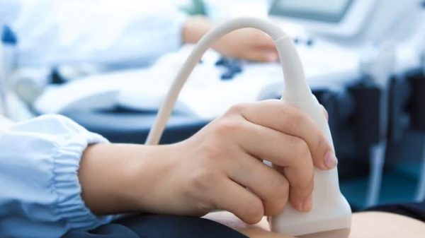
Real-time ultrasound is more sensitive than any other diagnostic test for ascites. More so, it is by far the easiest and cheapest method. It can detect ascitic fluid even if it is as low as 5 ml to 10 ml. Routine ultrasonography can visualize small quantities of ascitic fluid.
When you check an ultrasound result, uncomplicated ascites usually has a different appearance form complicated ascites. The appearance of uncomplicated ascites is homogeneous. More so, it appears to be a freely mobile collection with no echo.
There is also a demonstration of deep acoustic enhancement when ascites is uncomplicated. That’s not all. When you see uncomplicated ascites, it would not displace any organ. Rather, the fluid would lodge in-between organs.
An ultrasound would tell you a lot about ascites if it is present. But then, you don’t have to learn all of the signs. The doctors are trained to interpret the ultrasound results. So when they see it, they can tell a lot about your condition.
So then, ultrasound will not only help detect that ascitic fluid is present in your peritoneum. But it will also give lots of clues about the condition, its severity, and possibly its cause.
The thing with ultrasound is that the pattern changes with position. So then, your position while taking the ultrasound is very important.
The most sensitive position is the hand-knee position. The right-lateral decubitus position is very sensitive too. Your doctor and/or radiographer would guide you on how to take the right position for your ultrasound test.
A very sensitive position can even detect fluid volume as small as 100 ml. But then, there may be loculated collections. To confirm that the fluid is indeed ascitic, you will be asked to reposition quite a few times. If the ultrasonic patterns still suggest ascites, even with repositioning, then it is indeed ascites.
Differential Diagnosis of Ascites from GI Radiology
There are many reasons why ultrasound and other GI radiology tests can detect fluid in your peritoneum. So you should not jump to a conclusion if they detect some fluid. Wait till a consultant radiologist interprets the test and tells you the diagnosis.
Fluid volume is a woman’s peritoneum usually fluctuates as the goes through her monthly cycle. So there may be a physiological accumulation of fluid at some point. More so, young ladies sometimes have some fluid in their pelvis, and that is normal too.
Other possible causes of fluid accumulation include choleperitoneum, where bile leaks. This causes a type of ascites known as chylous ascites. There is also another type called pancreatic ascites. Ascites can also occur as a result of bladder trauma.
Ascites ultrasound is very sensitive, so cheap, and a lot easier to perform. It will give you almost all the answers you seek about the fluid in your peritoneum.